

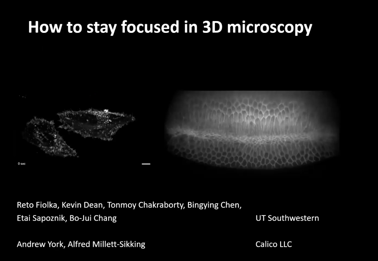
27th July 2020: 'How to stay focused in 3D microscopy' - Reto Fiolka,
Duration: 1 hour 8 mins
Share this media item:


Embed this media item:
Embed this media item:
About this item
| Description: |
Abstract: A conventional fluorescence microscope can directly form a three-dimensional image of a sample, however only within the focal plane it is sharp. Outside, aberrations and non-uniform magnification distort image formation. Over a decade ago, within the group of Professor Tony Wilson, it was realized that if certain conditions are met, a distortion and aberration free three-dimensional image can nevertheless be formed. This in itself is a surprise, and has spurred many applications based on this finding, ranging from remote focusing in raster scanning microscopy to light-sheet microscopes with a single objective interfacing the sample.
In this talk, I will present some of our most recent work that leverages this principle. I will first go over some of the theoretical aspects that ensure aberration free 3D image formation. I will then present a novel, rapid focusing technology that operates in an aberration free format. By transforming rapid lateral scanning into an aberration free axial scan motion, we can perform rapid two-photon microscopy. With an axial scan rate of 12 kHz and an effective numerical aperture (NA) of 1, we achieve high-speed, diffraction limited 3D imaging. Applications to neurological and cardiac imaging in model organisms will be presented. In the second part, I will present our latest work on high-resolution oblique plane microscopy (OPM). In OPM, a light-sheet is launched by a high NA objective at a shallow angle, and aberration free 3D imaging is leveraged to form a sharp image of the tilted light-sheet plane. OPM has traditionally partitioned the pupil of its primary objective into excitation and detection. As such, the spatial resolving power has often been modest, as only a subset of the primary objective’s NA was used. More recently, it has been realized that via careful engineering of the downstream optics, almost the full NA provided by the primary objective can be used for image formation, yielding increased resolving power and light-throughput. We show that such an OPM system can be used to image subcellular dynamics at high spatiotemporal resolution, combined with the convenience of traditional sample mounting. As such, we have used this microscope on samples that would be incompatible with conventional light-sheet systems, including multiwell plates and microfluidic channels. Bio: Reto Fiolka grew up in a small mountain town in Switzerland, and received a Master’s degree in Mechanical Engineering from ETH Zurich. He performed his PhD work in the laboratory of Professor Andreas Stemmer at ETH Zurich on structured illumination microscopy (SIM) and quantitative phase imaging. For his postdoctoral work, he went to HHMI’s Janelia Research Campus to work with the late Dr. Mats Gustafsson on multicolor 3D SIM. After the passing of Dr. Gustafsson, he performed secondary postdoctoral work under Dr. Meng Cui on phase conjugation and adaptive optics. In 2013, he was recruited to the Cell Biology Department at UT Southwestern by Dr. Sandra Schmid and Gaudenz Danuser, in an effort to bring advanced fluorescence microscopy to the campus. Since 2016, Reto Fiolka leads his own lab that is focused on 3D imaging technologies that push the boundaries of spatio-temporal resolution and optical penetration depth. The resulting microscopes are used in collaborative research programs across UT Southwestern. Reto Fiolka is grateful to the team led by Dr. Kevin Dean, which plays a crucial part in disseminating and applying the new technologies to cutting edge biomedical research problems. |
|---|
| Created: | 2020-07-27 17:12 |
|---|---|
| Collection: | Imaging ONE WORLD |
| Publisher: | University of Cambridge |
| Copyright: | Reto Fiolka |
| Language: | eng (English) |
| Distribution: |
World
|
| Keywords: | microscopy; lightsheet; |
| Explicit content: | No |
| Aspect Ratio: | 16:9 |
| Screencast: | No |
| Bumper: | UCS Default |
| Trailer: | UCS Default |
Available Formats
| Format | Quality | Bitrate | Size | |||
|---|---|---|---|---|---|---|
| MPEG-4 Video | 1280x720 | 2.57 Mbits/sec | 1.28 GB | View | ||
| MPEG-4 Video | 640x360 | 730.47 kbits/sec | 363.81 MB | View | ||
| WebM | 1280x720 | 950.75 kbits/sec | 473.52 MB | View | ||
| WebM | 640x360 | 295.34 kbits/sec | 147.09 MB | View | ||
| iPod Video | 480x270 | 484.08 kbits/sec | 241.10 MB | View | ||
| MP3 | 44100 Hz | 252.92 kbits/sec | 124.12 MB | Listen | ||
| Auto * | (Allows browser to choose a format it supports) | |||||