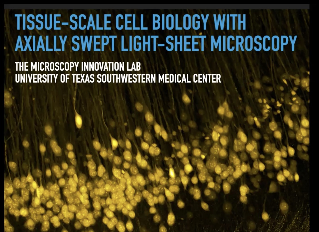
20th July 2020: 'Tissue-Scale Cell Biology with Axially Swept Light-Sheet Microscopy' - Kevin Dean,
Duration: 1 hour 14 mins
Share this media item:
Embed this media item:
Embed this media item:
About this item

| Description: |
Title: Tissue-Scale Cell Biology with Axially Swept Light-Sheet Microscopy
Speaker: Kevin Dean (UT Southwestern Medical Center, Dallas, Texas, USA) www.utsouthwestern.edu/research/core-facilities/microscopy-innovation/ Abstract: Major interdisciplinary efforts aim to comprehensively catalogue cellular diversity in a range of mouse and human tissue types. In an effort to support such scientific endeavors, we developed multiple light-sheet microscopes that deliver isotropic sub-cellular resolution throughout millimeters of tissue. These systems, collectively referred to as cleared-tissue Axially Swept Light-Sheet Microscopy (ctASLM), combine refractive index-independent illumination and detection optics, high-speed aberration-free remote focusing, and synchronous camera-based readout to maximize the field of view, optical sectioning, and axial resolution. The first variant of ctASLM is optimized for imaging large specimens such as an intact mouse brain with synapse-level detail, and provides a field of view 870 mm2 and an isotropic resolution of ~670 nm. The second variant is optimized for imaging subcellular detail deep within chemically cleared tissue sections, and provides a field of view of 330 mm2 and an isotropic resolution of ~285 nm. Both systems are orders of magnitude faster than a confocal microscope, and the high resolution variant delivers best-in-class axial resolution without necessitating multiview deconvolution, structured illumination, stimulated emission, or localization techniques. Owing to the performance of these microscopes, we successfully resolved and automatically segmented dendritic spines, evaluated the cellular composition of glomeruli throughout a kidney, visualized hematopoietic stem cell interactions in a bone marrow plug, quantitatively evaluated single cell morphology, and more. Thus, ctASLM not only provides global tissue architectures, but quantitatively detailed morphological and biochemical descriptions of the individual cells that compose tissues in health and disease. Bio: Kevin Dean was raised in a small town in Northern California, received his B.A. in Chemistry at Willamette University in Oregon, and was recognized twice as an ESPN Regional Academic All-American Running Back in Football. After college, he and two friends rode bicycles across the United States to raise money and awareness for Amyotrophic Lateral Sclerosis, which is more commonly known as Lou Gehrig’s Disease, in honor of one of his close friends that succumbed to the disease. He then received his Ph.D. in Biochemistry at the University of Colorado under the guidance of Dr. Amy Palmer and Dr. Ralph Jimenez. Here, his work focused on spectroscopy, protein engineering, and multi-parameter high-throughput microfluidic analyses and cell sorting. Following graduation, he established the first campus-wide light microscopy facility at the BioFrontiers Institute at the University of Colorado. Thereafter, he moved to the University of Texas Southwestern Medical Center in Dallas to perform his postdoctoral research under the guidance of Dr. Gaudenz Danuser and Dr. Reto Fiolka. During this time, he was named a Ruth L. Kirschstein Postdoctoral Fellow, received the Dean’s Discretionary Award at UT Southwestern, and was the runner-up for the UT Southwestern Brown-Goldstein Excellence in Postdoctoral Research award. Today, he runs a collaborative lab at UT Southwestern that brings cutting-edge computer vision and microscopy to biologists in an effort to advance our understanding of biological systems. He has been happily married for over a year now and is excited to announce that he has a son on the way. |
|---|
| Created: | 2020-07-20 17:26 |
|---|---|
| Collection: | Imaging ONE WORLD |
| Publisher: | University of Cambridge |
| Copyright: | Kevin Dean |
| Language: | eng (English) |
| Distribution: |
World
|
| Keywords: | microscopy; lightsheet; |
| Explicit content: | No |
| Aspect Ratio: | 16:9 |
| Screencast: | No |
| Bumper: | UCS Default |
| Trailer: | UCS Default |
Available Formats
| Format | Quality | Bitrate | Size | |||
|---|---|---|---|---|---|---|
| MPEG-4 Video | 1280x720 | 2.89 Mbits/sec | 1.57 GB | View | ||
| MPEG-4 Video | 640x360 | 917.79 kbits/sec | 497.44 MB | View | ||
| WebM | 1280x720 | 1.29 Mbits/sec | 718.17 MB | View | ||
| WebM | 640x360 | 372.71 kbits/sec | 202.01 MB | View | ||
| MP3 | 44100 Hz | 251.51 kbits/sec | 136.32 MB | Listen | ||
| Auto * | (Allows browser to choose a format it supports) | |||||

