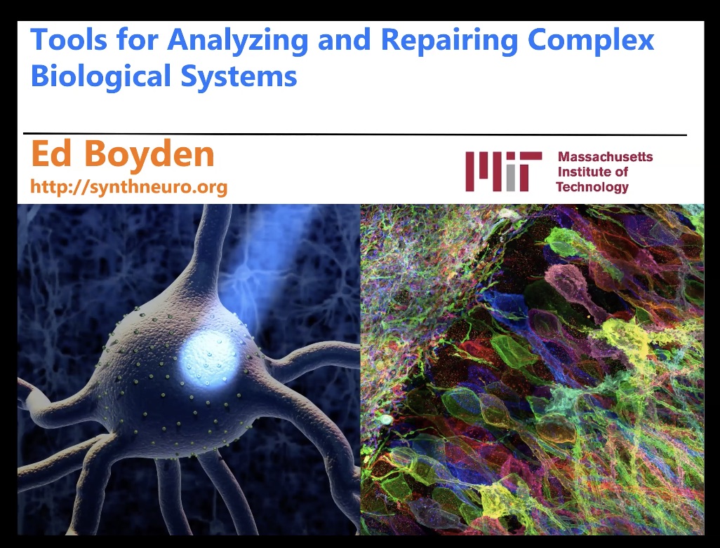
15th June 2020: 'Expansion Microscopy' - Ed Boyden, MIT Medialab & McGovern Institute, Cambridge, MA, USA
Duration: 55 mins
Share this media item:
Embed this media item:
Embed this media item:
About this item

| Description: |
Ed Boyden, PhD is Professor of Biological Engineering and Brain and Cognitive Sciences, MIT Media Lab and McGovern Institute’s Y. Eva Tan Professor in Neurotechnology
His groups’ research topics include: Tools for mapping, analyzing, and controlling brain computations, New therapeutics for neurology and psychiatry, Approaches for ground-truth understanding of living systems Abstract: Recently our group developed a new method for imaging, with nanoscale spatial precision, the identity and location of proteins, nucleic acids, and other kinds of biomolecules in 3-D throughout intact cells and tissues, by physically expanding such samples in an isotropic fashion. By embedding biological specimens in a swellable polymer, anchoring key biomolecules or labels to the polymer, mechanically homogenizing the specimens, and then swelling the samples isotropically (Science 347(6221):543-8), we found that it was possible to physically swell samples by ~4.5x in linear dimension, enabling nanoscale imaging of complex biological systems in 3-D –- on ordinary diffraction-limited microscopes. Since then, we have been working to improve the expansion factor further, aiming for ~10x-100x expansion in linear dimension, which would ideally give an effective resolution in the nanometers to low tens of nanometers. We have also been working to incorporate multiplexed imaging of biomolecules into the expansion microscopy context. We have also developed protocols for expansion microscopy of paraffin-embedded, fresh frozen, and other kinds of pathology specimens. Our hope is that this toolset will enable the powerful, democratized identification and localization of perhaps thousands of different kinds of biomolecule, with nanoscale precision, throughout cells and tissues- a key step towards understanding the configuration of complex biological systems in normal and disease states. Bio: http://syntheticneurobiology.org/people/display/71/11 Selected Publications: D., Oran*, S. G. Rodriques*, R. Gao, S. Asano, M. A. Skylar-Scott, F. Chen, P. W. Tillberg, A. H. Marblestone**, and E. S. Boyden**. “3D nanofabrication by volumetric deposition and controlled shrinkage of patterned scaffolds.” Science 362, no. 6420 (2018): 1281-1285 (*, equal contributors; **, equal contributors) Tillberg, P.W.*, Chen F.*, Piatkevich K.D., Zhao Y., Yu C.-C., English B.P., Gao L., Martorell A., Suk H.-J., Yoshida F et al. “Protein-retention expansion microscopy of cells and tissues labeled using standard fluorescent proteins and antibodies.” Nature Biotechnology 34 (2016): 987-992 (*, co-first authors) . Chen, F.*, Tillberg P.W.*, and Boyden E.S. “Expansion Microscopy.” Science 347, no. 6221 (2015): 543-548 (*, equal contribution) . Image: an expanded piece of the brain, which is a zoom-in into Fig. 3f from, http://syntheticneurobiology.org/publications/publicationdetail/251/25 |
|---|
| Created: | 2020-06-25 17:29 |
|---|---|
| Collection: | Imaging ONE WORLD |
| Publisher: | University of Cambridge |
| Copyright: | Ed Boyden |
| Language: | eng (English) |
| Distribution: |
World
|
| Keywords: | Expansion; microscopy; |
| Explicit content: | No |
| Aspect Ratio: | 16:9 |
| Screencast: | No |
| Bumper: | UCS Default |
| Trailer: | UCS Default |
Available Formats
| Format | Quality | Bitrate | Size | |||
|---|---|---|---|---|---|---|
| MPEG-4 Video | 1280x720 | 2.94 Mbits/sec | 1.19 GB | View | ||
| MPEG-4 Video | 640x360 | 1.65 Mbits/sec | 684.60 MB | View | ||
| WebM | 1280x720 | 2.67 Mbits/sec | 1.08 GB | View | ||
| WebM | 640x360 | 752.0 kbits/sec | 303.02 MB | View | ||
| iPod Video | 480x270 | 481.51 kbits/sec | 193.97 MB | View | ||
| MP3 | 44100 Hz | 249.76 kbits/sec | 100.73 MB | Listen | ||
| Auto * | (Allows browser to choose a format it supports) | |||||

